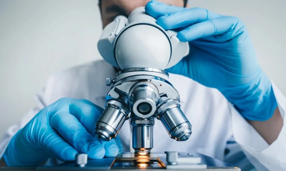
Low-power optics lie somewhere between the naked eye and complete microscope capabilities. When it comes to examining any solid pieces, assemblies, or living samples, getting the proper configuration may be crucial to spotting a minute defect. Making such an assessment involves taking into account the optical path, lighting conditions, and rig stability in operation.
Different types of precision jobs don’t have the same optical method of magnification, which depends on the depth of field and resolution required. In certain cases, technicians get confused between a stereo microscope vs dissecting microscope based on some differences in their designs, working distances, and illumination systems.
Differences in Optical Design
The split comes down to how the optics are built. A basic dissecting scope usually runs on a single shared objective, or a simplified Greenough path, enough for straightforward handling. Higher-end stereo setups use a Common Main Objective (CMO) design with parallel light paths, keeping the image distortion-free – useful for reticles or a mounted camera.
For more on optical path design, the National Institute of Standards and Technology publishes guidance on optical metrology.
Magnification Ranges and Working Distance
Most low-power optics fall between 2x and 300x. With an increase in magnification, working distance and depth of field reduce – the clearance below the lens becomes important during tool manipulation while assembling or dissecting.
| Microscope Series | Magnification Range | Working Distance Range | Depth of Field | Mostly Used For |
|---|---|---|---|---|
| Simple Dissecting Series | 5x – 40x | 70mm – 120mm | High | General-use tissue dissection, introductory microscopes |
| Greenough Series | 10x – 100x | 80mm – 100mm | Moderate | Soldering PCBs, quality control, general use |
| Parallel (CMO) Series | 8x – 300x | 30mm – 160mm (modular) | Adjustable | Metrology and precision assembly |
Small numerical apertures mean greater depth of field, which will keep uneven specimens sharp rather than becoming out of focus.
Lighting Systems and Reflection Control
Illumination is what makes contrast possible on opaque materials – a plain ring light can flood shiny metal or silicon in glare, burying hairline cracks. Segmented LED rings, polarizing filters, and oblique lighting fix this:
- Polarized light: reduces glare from polished metals and glass.
- Oblique lighting: casts shadows at an angle, showing surface details.
- Transmitted bases: light translucent samples from below.
Research related to illumination and contrast has been published by the National Institutes of Health (NIH).
Ergonomics and Long-Session Usability
Standard eyepieces force a fixed head position, adding up to neck and eye strain over a shift. Eyepiece-less displays and digital heads widen the exit pupil to 30mm, letting operators shift position freely, even with glasses on, keeping focus sharper and cutting mistakes. Articulated boom stands add stability, isolating the scope from vibration on a busy floor.
Choosing Optics for Specific Applications
- Medical Device Assembly: needs strong depth of field, cleanroom-ready hardware, and compliant documentation.
- Electronics Repair: needs generous working distance (100mm-plus) for a soldering iron to move underneath.
- Biological Research: needs adjustable lighting for living tissue without heat damage.
An understanding of how the stereo microscope vs dissecting microscope design manages depth ensures the equipment is suitable for the task. The Occupational Safety and Health Administration archives the lab equipment safety standards.
Strategic Optical Selection
Selecting low power lenses involves a balance among resolving power, workspace, and user ergonomics. The choice of either an entry-level dissection microscope or a complete parallel stereo system is based on the need for precision and quantity of work done. Matching hardware to actual demands protects workflow, cuts fatigue, and keeps inspection accuracy consistent.

Comments are closed.